Preclinical: Prime Editing Restores...
CMN Special
Oct. 7, 2026

Retinitis pigmentosa progressively destroys rod photoreceptors and is followed by secondary cone loss. Around 30%–40% of cases are autosomal dominant, and mutations in RHO, which encodes rhodopsin, are a major cause. Many of the more than 150 known RHO variants have dominant-negative or gain-of-function effects. Adding a normal gene copy may therefore be insufficient: the harmful allele must also be silenced or replaced. Mutation-specific editing is possible, but each variant may require its own guide RNA.
“Our findings highlight the critical importance of HITI donor DNA design in determining productive genome-editing outcomes”Esposito et al.
Federica Esposito and colleagues instead developed a strategy intended to work independently of the particular pathogenic RHO variant. Published recently in Cell Reports Medicine, it combines CRISPR-Cas9 with homology-independent targeted integration, or HITI, which uses non-homologous end joining to insert donor DNA at a programmed double-strand break. Two AAV2/8 vectors were injected beneath the retina. One expressed Streptococcus pyogenes Cas9 specifically in photoreceptors; the other carried a guide RNA targeting intron 1 of human RHO and a promoterless donor cassette containing wild-type RHO coding sequence.
Cas9 cuts the genomic target and releases the donor cassette from its vector. Cellular repair can then join the donor to intron 1, disrupting expression of the targeted allele while placing functional RHO under the endogenous promoter (see Figure 1). This is not correction of the P23H nucleotide. It is targeted gene insertion coupled to functional inactivation of the existing allele, designed to preserve physiological regulation of rhodopsin and avoid a separate treatment for every dominant RHO mutation.
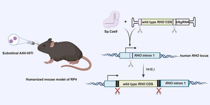
The first donor contained three stop codons intended to block mutant rhodopsin production. Integration was detectable, but expression was poor because the premature stops promoted nonsense-mediated mRNA decay. The researchers replaced them with a CL1 degron linked to a furin site and P2A peptide. This directs the truncated endogenous rhodopsin fragment towards degradation while allowing production of wild-type rhodopsin and an eGFP reporter from the same transcript. As the authors note, “Our findings highlight the critical importance of HITI donor DNA design in determining productive genome-editing outcomes.”
The optimised system was tested in mice carrying one human RHO allele with the P23H mutation and one normal mouse Rho allele. At one month, approximately 10% ± 4% of rods in the AAV-transduced outer nuclear layer expressed eGFP, indicating productive integration. The value remained about 10% ± 6% after one year. PCR-free nanopore sequencing of the injected temporal retina found donor integration in approximately 11% of analysed human RHO alleles. These estimates use different denominators and should not be combined.
“Together, these findings demonstrate durable structural, functional, and vision-related behavioral improvements following AAV-HITI treatment”Esposito et al.
Editing outcomes were heterogeneous. Among integration events recovered by long-range PCR and nanopore sequencing, about 43% were full-length donor insertions in the intended orientation, while 19% were reverse integrations and 31% were partial integrations; smaller fractions contained other AAV-derived sequences. Separately, short-read sequencing detected insertions or deletions at the Cas9 cut site in approximately 18% of reads, against 0.04% in scramble-guide controls. These changes may have contributed to benefit by disrupting mutant P23H expression, but they also show that HITI does not generate one uniform repair product.
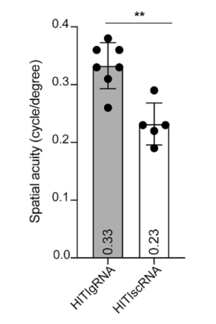
Despite mosaic editing, treated eyes performed better than contralateral eyes receiving the same vectors with a scramble guide. Electroretinographic responses were higher, and optical coherence tomography showed better preservation of the photoreceptor layer. Mutant RHO-P23H-RFP fluorescence was reduced approximately threefold in the temporal retina. At one year, optokinetic testing showed a spatial acuity of 0.33 cycles/degree in edited eyes, compared with 0.23 in controls and approximately 0.36 in wild-type mice (see Figure 2). “Together, these findings demonstrate durable structural, functional, and vision-related behavioral improvements following AAV-HITI treatment,” the researchers conclude. Correcting a minority of rods could therefore produce a measurable benefit across the treated region.
The safety analyses identify issues requiring further study. Sequencing revealed unintended Cas9 activity at intronic sites in UTP6 and JARID2 in human HEK293 cells, as well as low-frequency integration of donor or Cas9-vector sequences. No aberrant UTP6 or JARID2 transcripts were detected, but kidney-derived HEK293 cells do not reproduce the chromatin or DNA-repair biology of mature human rods. The authors could not obtain sufficiently transduced human retinal organoids for reliable off-target analysis.
The mouse model also answers only part of the translational question. Its human-specific guide targeted the mutant human RHO allele but not the normal mouse Rho allele. In patients, the target sequence is present in both human alleles, so productive and non-productive events could affect the healthy copy and disturb rhodopsin dosage. Evaluation in a fully humanised model or non-human primates will therefore be needed to establish the balance between efficacy and genomic risk. The study demonstrates durable proof of concept for partial, mutation-agnostic editing, not complete or clinically validated correction.
The study was led by Federica Esposito and Alberto Auricchio at the Telethon Institute of Genetics and Medicine in Pozzuoli, Italy. It was published in Cell Reports Medicine on 11 September 2026.
To get more CRISPR Medicine News delivered to your inbox, sign up to the free weekly CMN Newsletter here.
ArticleCMN HighlightsNewsStickyIn vivoMicroinjectionAdeno-associated virus (AAV)Retinitis PigmentosaEyeCas9Pre-clinical